Shape analysis of the nasal complex among South African groups from CBCT scans
DOI:
https://doi.org/10.17159/sajs.2024/12972Keywords:
human variation of South African groups, cone-beam computer tomography scans, geometric morphometric methods, South African standard facial reconstruction methodsAbstract
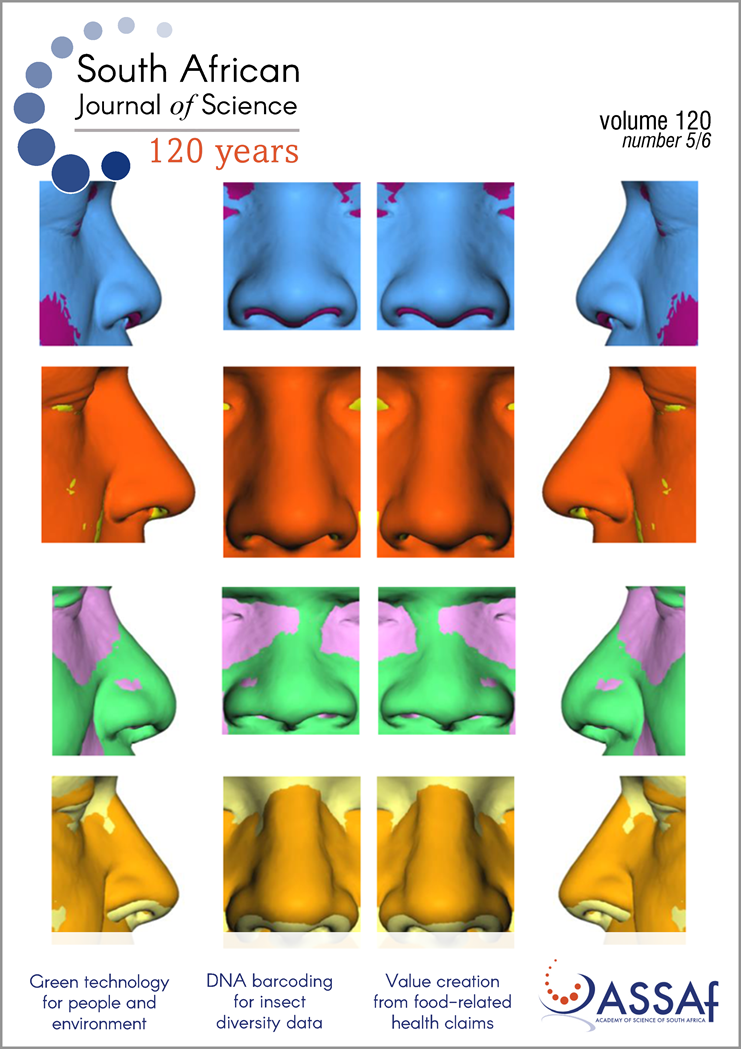
Three-dimensional (3D) anatomical extraction techniques could help the forensic anthropologist in a precise and inclusive assessment of biological phenotypes for the development of facial reconstruction methods. In this research, the nose morphology and the underlying hard tissue of two South African populations were studied. To this end, a 3D computer-assisted approach based on an automated landmarking workflow was used to generate relevant 3D anatomical components, and shape discrepancies were investigated using a data set of 200 cone-beam computer tomography (CBCT) scans. The anatomical landmarks were placed on the external nose and the mid-facial skeleton (the nasal bones, the anterior nasal aperture, the zygoma, and the maxilla). Shape differences related to population affinity, sex, age, and size were statistically evaluated and visualised using geometric morphometric methods. Population affinity, sexual dimorphism, age, and size affect the nasal complex morphology. Shape variation in the mid-facial region was significantly influenced by population affinity, emphasising that shape variability was specific to the two population groups, along with the expression of sexual dimorphism and the effect of ageing. In addition, nasal complex shape and correlations vary greatly between white and black South Africans, highlighting a need for reliable population-specific 3D statistical nose prediction algorithms.
Significance:
- 3D anatomical structures were acquired and extracted from 200 CBCT scans of modern South Africans.
- Geometric morphometric methods were applied.
- Soft- and hard-tissue nasal complex morphology vary across South African groups.
Additional Files
Published
Issue
Section
License

All articles are published under a Creative Commons Attribution 4.0 International Licence
Copyright is retained by the authors. Readers are welcome to reproduce, share and adapt the content without permission provided the source is attributed.
Disclaimer: The publisher and editors accept no responsibility for statements made by the authors
How to Cite
- Abstract 578
- PDF 115
- EPUB 441
- XML 394
- Peer review history 27






.png)